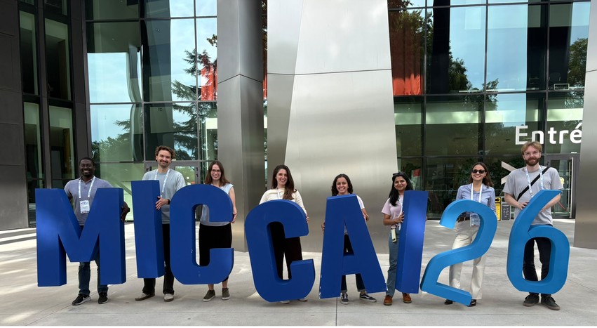
Hertie AI at MICCAI 2026
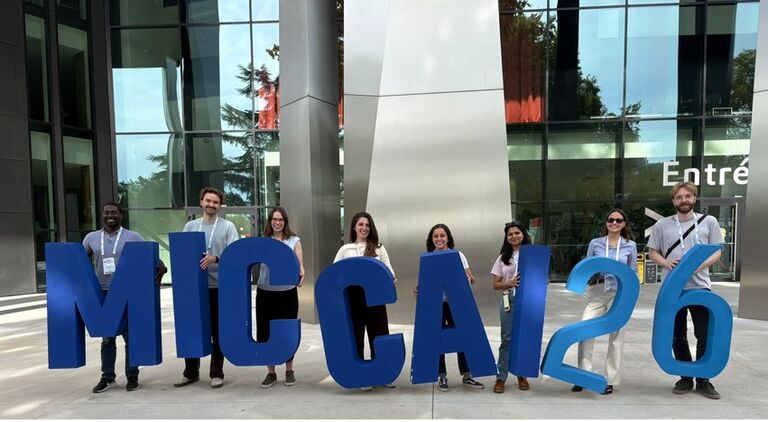
Hertie AI at MICCAI 2026
From 27 September to 1 October, scientists from the Hertie Institute for AI in Brain Health (Hertie AI) attended the 29th International Conference on Medical Image Computing and Computer-Assisted Intervention (MICCAI 2026) in Straßburg, France. Hertie AI contributed two papers to the main conference, one paper to the iMIMIC workshop, and helped organise the BraTS Inpainting Challenge. These activities spanned retinal imaging, brain recordings, and image synthesis for brain tumour research.
Samuel Ofosu Mensah presented “Towards Interpretable Foundation Models for Retinal Fundus Images”, jointly led with co-first author Camila Roa, through both an oral and a poster presentation at the workshop on Interpretability of Machine Intelligence in Medical Image Computing (iMIMIC). The work introduces Dual-IFM, a model designed to make its predictions easier to understand. It highlights the regions of a retinal photograph that contribute to a prediction and provides a two-dimensional map for exploring patterns across images. Trained on more than 800,000 retinal photographs, it achieved performance comparable to a much larger foundation model in the evaluated tasks.
At the main conference, Meghal Dani’s paper, “Parameter-Efficient Self-Supervised Adaptation for EEG-FM under Fixed Computational Budgets,” investigated how pretrained models for electroencephalography (EEG) can be adapted to new clinical datasets with limited computing resources. The study showed that updating a small portion of a model using unlabelled recordings improved performance across tasks including seizure and abnormality detection, offering insights into more efficient use of EEG foundation models.
Gustav Schmidt and Sarah Müller, presented “Ordinal Diffusion Models for Color Fundus Images.” Their approach generates retinal photographs while explicitly accounting for the ordering of diabetic retinopathy severity. This allows smooth transitions between disease stages and produced images with improved visual realism and closer agreement with the intended severity compared with a model treating each stage as a separate category.
Paula Buzduga, Erik Großkopf and Florian Kofler also co-organised the BraTS 2026 Inpainting Challenge, part of the wider Brain TumorS challenge programme. Participants developed methods to fill masked regions of brain MRI scans with plausible healthy tissue – a process known as inpainting. The challenge provides a common benchmark for comparing these methods, supporting research into how images affected by brain tumours can be analysed using tools designed for healthy brain anatomy.
Together, these contributions reflect complementary directions in our research: making medical AI easier to interpret, adapting it under practical resource constraints, incorporating disease structure into image generation, and supporting shared evaluation through community challenges. MICCAI provided an international forum for sharing this work with the wider medical computing community.